

A team of scientists, including Dr. hab. Karolina Mikulska-Rumińska from the Department of Biophysics at the Institute of Physics, Nicolaus Copernicus University in Toruń, has made a breakthrough discovery related to the process of ferroptosis, revealing a previously unknown role of plasmalogens, a class of ether phospholipids, in the regulation of ferroptotic cell death. The results of the study were published in the prestigious journal Nature Communications.
In the human body, cells are constantly formed, perform specific functions, and die, while their number remains relatively stable thanks to precise regulatory mechanisms. This dynamic balance allows tissues to maintain their proper structure and function. An important component of this balance, essential for maintaining homeostasis, is regulated cell death.
Ferroptosis is a form of regulated cell death characterized by an increasing level of lipid hydroperoxides in cellular membranes, associated with the presence of reactive iron and weakened antioxidant defenses of the cell. In this process, reactive oxygen species initiate lipid oxidation, leading to damage to cellular membranes, as well as proteins and genetic material, ultimately resulting in cell death. Ferroptosis is being intensively studied as a potential mechanism that could be exploited in cancer therapy, while its excessive activation may contribute to tissue damage, including in brain injury, kidney diseases, and asthma. Ferroptosis has also been linked to many pathological conditions, including neurodegenerative diseases, sepsis, brain trauma, and UV-induced skin damage. Although ferroptosis attracts considerable scientific interest, it still remains an incompletely understood process.
The team, which includes Dr. hab. Karolina Mikulska-Rumińska, has been collaborating for nearly ten years, intensively exploring various aspects of ferroptosis. During this time, its members have published several important studies on ferroptosis and regulated cell death, including in Cell, Journal of Clinical Investigation, Angewandte Chemie International Edition, JACS, PNAS, Nature Metabolism, and Nature Chemical Biology. The team consists of several independent experimental and theoretical groups representing different scientific disciplines, including chemistry, physics, biology, and medicine.
In the latest study, entitled “15-LOX-catalytic bias towards ether-(alkenyl)-ETE-PEs oxidation bestows selectivity of PRO-ferroptotic cell death signaling”, published on June 17, 2026 in Nature Communications, the authors revealed a new mechanism regulating ferroptosis. The study demonstrated that specific ether lipids, namely plasmalogen phosphatidylethanolamines containing a polyunsaturated fatty acid chain, are preferentially oxidized by enzymes from the 15-lipoxygenase family (Fig. 1). The lipid products formed in this process act as pro-ferroptotic signals and may contribute to cellular damage in inflammatory diseases, cancer, and tissue injuries.
The findings challenge the simplified view that widely occurring plasmalogens function in cells exclusively as protective antioxidants. The authors showed that, in a specific biochemical context, the same lipids can become a source of signals leading to cell death. The published research combined redox lipidomics, biochemical and cellular experiments, disease models, and molecular dynamics simulations, which helped explain why the 15-LOX enzyme selectively oxidizes this particular class of lipids.
The discovery is important for understanding the molecular basis of ferroptosis and diseases in which uncontrolled lipid oxidation plays a significant role, such as asthma, cancer, brain injury, and skin damage. The mechanism described in the publication may point to new therapeutic targets related to inhibiting the selective oxidation of lipids by 15-lipoxygenases.
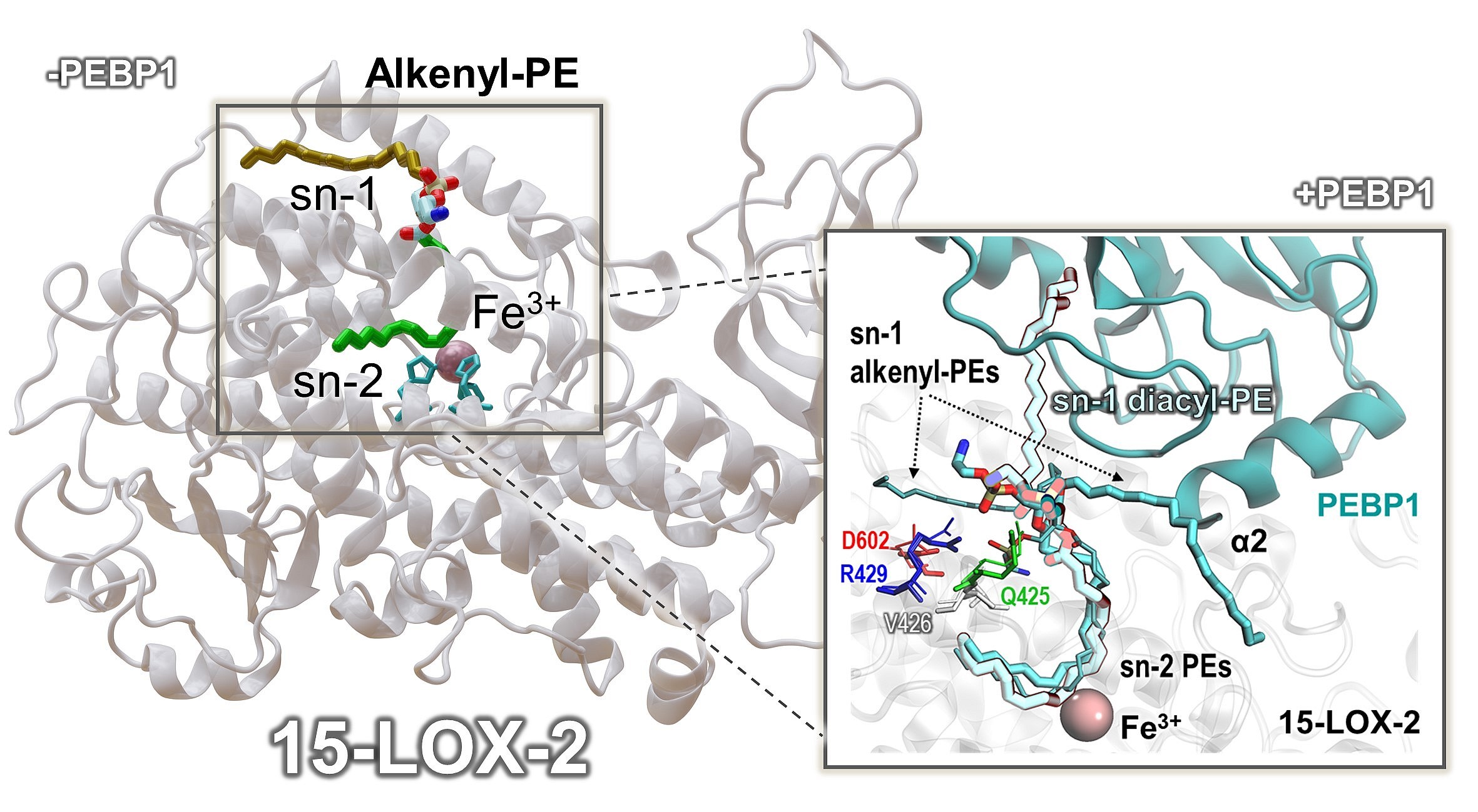
Figure 1. Structural model of the 15-lipoxygenase-2 complex with plasmalogen PE and comparison with PEBP1-stabilized diacyl-PE lipids. The left panel shows the three-dimensional structure of 15-LOX-2 bound to plasmalogen phosphatidylethanolamine (alkenyl-PE), with the sn-1 chain shown in brown and the oxidizable sn-2 chain shown in green. The lipid is positioned near the catalytic center of the enzyme, which contains an Fe³⁺ ion coordinated by histidine residues and the C-terminus of the protein. The inset shows the substrate arrangement within the catalytic pocket of 15-LOX-2 and compares the orientation of alkenyl-PE and diacyl-PE lipids in the presence of PEBP1. The highlighted amino acids illustrate interactions responsible for the distinct positioning of the sn-1 chain in diacyl-PE and alkenyl-PE lipids. In the case of diacyl-PE, which the group identified as pro-ferroptotic lipids in a 2017 study published in Cell, PEBP1 stabilizes a productive lipid orientation that promotes oxidation of the sn-2 chain. In contrast, in plasmalogen PE, due to the presence of an ether linkage at the sn-1 position, the sn-1 chain does not form stable contacts with PEBP1, and oxidation by 15-LOX-2 occurs independently of this protein. These findings indicate that diacyl-PE and alkenyl-PE may represent two classes of pro-ferroptotic lipids involved in ferroptosis through distinct molecular mechanisms. The inset is taken from the Supplementary Information of the above-mentioned publication in Nature Communications.